Tracking cancer-fighting immune cells with advanced MRI technology
Dr. Fanny Chapelin's team used fluorine-based MRI to follow macrophages gathering in mouse breast and colon tumours after radiation, revealing different immune responses between the cancer types.


A groundbreaking study introduces a new method to track the body’s immune response during cancer treatments using magnetic resonance imaging, potentially transforming how we understand tumor development and treatment success. This research, led by Dr. Fanny Chapelin, Harrison Yang and Brock Howerton from the University of Kentucky and the University of California San Diego, shows how magnetic resonance imaging can be used to observe macrophages—immune cells that are crucial in how tumors respond to radiation therapy. Published in Cancers, the study offers fresh insights that could lead to more effective cancer treatments by providing real-time images of tumor changes during therapy.
“Radiation therapy has long been used to treat cancer, but it has been challenging to track the body’s immune response during the process,” explained Dr. Chapelin. “Our study demonstrates how magnetic resonance imaging can monitor macrophage activity inside tumors without needing to perform invasive procedures, giving us a clearer view of the tumor environment.”
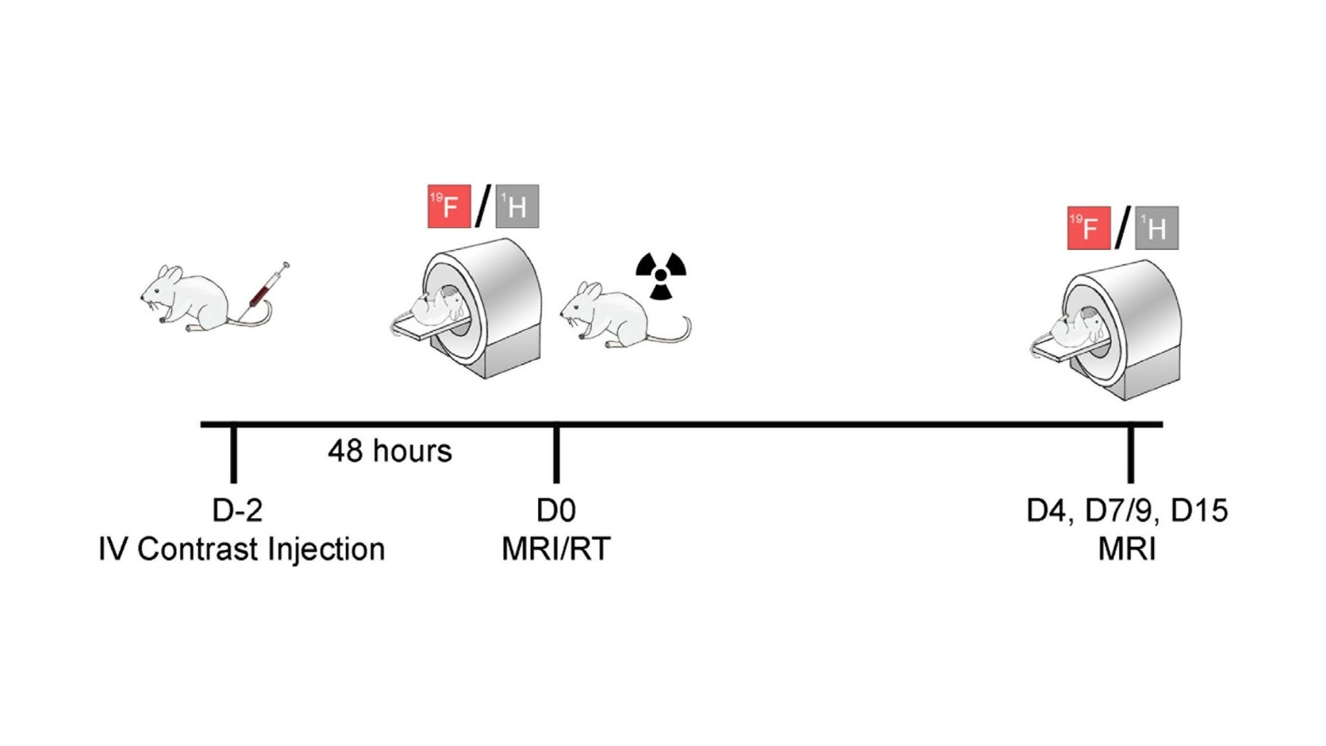
The researchers used a special fluorine-based compound, injected into mice with breast or colon cancer, to label macrophages, allowing them to be seen in magnetic resonance imaging scans. “This allowed us to follow the movement and behavior of these immune cells in real time after radiation therapy,” Yang shared. The study found that macrophages, which are often involved in fighting infections, were drawn to the tumors after treatment. However, the way the immune cells reacted differed between breast and colon cancers.
Fluorine-based magnetic resonance imaging provides a noticeable advantage. This approach offers a clean, quantifiable signal, unlike traditional magnetic resonance imaging, which often uses more complex agents to make things visible. With this method, scientists can observe how macrophages behave as the immune system reacts to the therapy.
In the case of colon cancer, tumors that were treated with radiation showed a dramatic increase in macrophages, which stopped the tumor from growing within a little over a week. Meanwhile, tumors that were not treated continued to grow aggressively. “We noticed a significant boost in the fluorine signal, which showed us that more macrophages were moving into the treated tumors,” Dr. Chapelin said. This increased immune activity seemed to directly link to the shrinking of the tumor, showcasing how crucial the immune system is in the fight against cancer following radiation.
In contrast, the breast cancer model showed a slower response from the immune cells, but there was still noticeable shrinkage of the tumor after radiation. “Although the breast tumors didn’t stop growing as fast as the colon tumors, we did observe a moderate rise in macrophage activity and a steady reduction in tumor size over time,” noted Howerton.
These results are particularly important because they suggest that the activity of macrophages, as shown by magnetic resonance imaging, could act as a sign of how well the treatment is working. This could help doctors assess the progress of radiation therapy without needing to perform biopsies, which can be invasive and uncomfortable for patients. “This method offers a real-time, non-invasive way to observe how the body is responding to cancer treatment, which is a significant step toward more personalized care for patients,” Dr. Chapelin added.
The potential applications of this method go beyond just tracking macrophages. By giving doctors a clearer picture of the tumor’s internal environment, it could help predict if the cancer might return, which is a common concern after treatment. Macrophages inside tumors can either help fight cancer or, in some cases, encourage its growth. Therefore, being able to monitor these immune cells in real time could give doctors valuable clues about the likelihood of the cancer coming back.
The research team also noted that this technology could be useful for other cancer treatments, including those that boost the immune system’s ability to fight the disease. “By understanding how immune cells like macrophages respond to different therapies, we could eventually tailor treatments to each patient, making them more effective,” Dr. Chapelin explained.
This breakthrough has the potential to revolutionize cancer treatment by offering a non-invasive way to monitor the immune response, avoiding the need for frequent and uncomfortable procedures like biopsies. The researchers plan to continue exploring how this technology can be used in different cancer types and aim to bring it into clinical practice. “Our goal is to move this technology from the lab to the clinic, where it can give doctors real-time feedback and help improve treatment outcomes for patients,” Dr. Chapelin said.
In conclusion, the use of magnetic resonance imaging to track macrophages during cancer treatment offers a promising new way to improve the effectiveness of radiation therapy. By providing a non-invasive method to monitor what’s happening inside the tumor, this technology could help doctors better understand how tumors are reacting to treatment, predict whether cancer might return, and tailor therapies to provide the best possible results for patients.
About the researchers

Fanny Chapelin
Fanny Chapelin is an Assistant Professor in the Departments of Bioengineering and Radiology at University of California San Diego. Fanny received her Ph.D. in Bioengineering from University of California, San Diego in 2019.… She then became the first woman faculty in the Department of Biomedical Engineering at University of Kentucky where she developed her research program for four years. She has been developing non-invasive imaging techniques to visualize cellular therapies and inflammation in cancer and other immune disorders. Her research findings have led to several publications, and their impact was recognized through numerous awards, including France’s Engineer of the Year Award for Science, a NIH KL2 award. She is a member of the International Society for Magnetic Resonance in Medicine, a Scialog Fellow and former UK Research Scholar. Her research aims to provide scientists and clinicians with ways to visualize cell distribution, fate and efficacy to improve clinical practice and patient care.

Harrison Yang
Harrison Yang is a senior at the University of Kentucky studying Biomedical Engineering under the Singletary Scholarship. During his undergraduate career, he has conducted research in cancer imaging and immunology laboratories. The Commonwealth Undergraduate Research Experience and Markey STRONG Scholars Program have funded his research endeavors.… His work has been recognized at the university level by the Sophomore Trailblazer in Biomedical Engineering Award and nationally by the Barry M. Goldwater Scholarship. He intends to continue researching cancer immunology following completion of his bachelor's degree.

Brock Howerton
Brock Howerton is a second-year Ph.D. graduate student in the Department of Bioengineering at the University of California, San Diego. He received his M.S. in Chemistry from the University of Kentucky, where he developed a strong interest in organic/polymer chemistry and drug delivery.… Currently, Brock is focused on developing novel immunoimaging contrast agents for cancer detection and T cell tracking using 19F/1H MRI. He is an active member of both the International Society for Magnetic Resonance in Medicine and the UCSD Bioengineering Graduate Society. His work centers on creating functional synthetic platforms that offer clinically translatable imaging tools. Brock's research aims to bridge the gap between laboratory discoveries and practical medical applications, providing clinicians with innovative tools to improve the diagnosis and treatment of diseases.
Journal reference
Yang, H., Howerton, B., Brown, L., Izumi, T., Cheek, D., Brandon, J.A., Marti, F., Gedaly, R., Adatorwovor, R., & Chapelin, F. “Magnetic Resonance Imaging of Macrophage Response to Radiation Therapy.” Cancers, 2023, 15, 5874. DOI: https://doi.org/10.3390/cancers15245874