Doctors Now Have a More Intelligent Way to Analyze Tissue in Cancer Patients
Dr. Sahil Saraf's team found that an AI tool for scoring a cell-growth stain in sarcoma samples made pathologists' assessments more consistent, working best alongside human review.


Researchers have developed a computer-based tool to improve the precision of Ki-67 testing, a method that examines a protein found in cells to determine how fast they are growing, an important marker used to assess cancer growth and progression. Led by Dr. Sahil Saraf, a team of scientists from Qritive Pte. Ltd. and Singapore General Hospital collaborated on this advancement. Their study has been published in the peer-reviewed journal Heliyon.
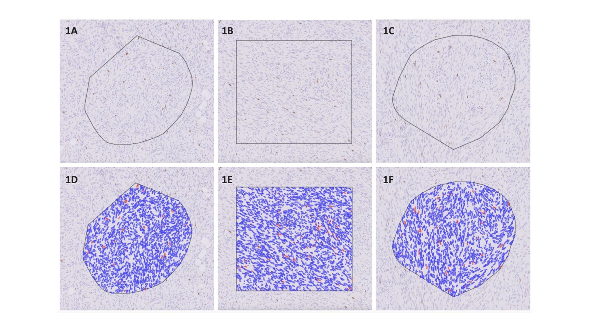
Measuring Ki-67 helps doctors understand how quickly cancer cells are multiplying. However, manually examining Ki-67-stained slides is time-consuming and can lead to inconsistencies among pathologists, medical specialists who study tissues and diagnose diseases. To improve this, the researchers designed a computer-driven system that automates the scoring process, making results more reliable and reducing the chance of human error. It was observed that the best results were seen when the AI and pathologist worked together. In their study, Dr. Saraf and colleagues analyzed numerous cases of sarcoma, a type of cancer that develops in bones and soft tissues, reviewing hundreds of tissue areas under the microscope. Doctors assessed Ki-67 levels both with and without computer assistance, showing that the tool greatly improved consistency between evaluations.
Results showed that using the computer-based system significantly reduced differences between pathologists’ assessments. “This tool enhances the accuracy and reliability of Ki-67 readings by reducing individual interpretation differences and standardizing the scoring method,” explained Dr. Saraf. Since Ki-67 is a nuclear stain, the system works by separating and identifying cell nuclei and categorizing them based on stain intensity through advanced image-processing techniques.
Doctors who initially assessed Ki-67 manually found that the computer-supported method led to more uniform results. “The findings indicate that computer assistance can provide essential support in pathology, helping to ensure more precise tumor classification and better treatment decisions,” noted Dr. Saraf. The study also found that doctors agreed with the computer’s results in the vast majority of cases, demonstrating strong alignment between human and automated assessments.
New technology is set to transform cancer diagnostics. By improving consistency and efficiency, this tool can help provide more accurate tumor profiling, a detailed analysis of a tumor’s characteristics to guide treatment, leading to better treatment options for patients. While human review remains vital, computer-assisted evaluations are proving to be a valuable addition to medical practice. Further improvements to the system are expected to enhance its ability to identify tumor areas with even greater accuracy, refining its diagnostic performance.
Dr. Saraf and his team’s findings highlight how technology can help improve medical testing, offering a reliable tool for pathologists to use in their work. As the healthcare field increasingly adopts digital solutions, innovations like this will become essential to modern cancer diagnosis, leading to better and more efficient patient care.
About the researchers

Dr. Sahil Saraf
Dr. Sahil Saraf is a seasoned pathologist with over 13 years of expertise in the field. He is known for his leadership, clinical skills, consulting capabilities and for playing a key role in advancing diagnostic techniques and research. Dr.… Saraf completed a Fellowship in Histopathology at the Singapore General Hospital and has held prominent positions, including his current designation as a senior specialist, Dept of Pathology at V.G. Saraf Memorial Hospital, Medical Director at Qritive PTE. LTD, and Consultant Pathologist at DDRC SRL Laboratory. Additionally, he serves as the director of two companies specializing in chemical manufacturing.A significant contributor to the integration of AI in healthcare, Dr. Saraf has developed diagnostic models for prostate grading and lymph node diagnoses. At Qritive, he was instrumental in securing essential certifications and helping the company gain recognition in renowned accelerator programs. Dr. Saraf’s work has been published in various medical journals, and he has been invited to present as a keynote speaker at medical conferences.In 2024, Dr. Saraf was chosen for a platform presentation at the USCAP annual conference in Baltimore, MD, where he presented his paper, "A Deep Learning Module to Assist Pathologists in Identifying Lymph Node Metastasis in Breast Tumor Resections." His research earned him the 1st place ISBP-BCRF Larry Norton MD Trainee Abstract Award in the Breast Pathology research category.Dr. Saraf completed his MD at Vydehi Institute of Medical Sciences and Research Center and his MBBS at JJM Medical College. He has also pursued several fellowships, internships, and continuing education programs.

Dr. Priti
Dr. Priti is Professor in Department of Pathology, at Lady Hardinge Medical College, New Delhi, India. She did her Undergraduate training from Medical College Calcutta, India and MD pathology from All India Institute of Medical Sciences, New Delhi, India. Her fields of interest are Surgical Pathology, Oncopathology, and Molecular Pathology.… She is a passionate academician and loves teaching undergraduate and postgraduate trainees. She has presented numerous scientific posters and oral papers for which she also has won National level Awards. She has authored many research publications in national and international journals.

Santosh KV
Santosh KV is a consultant pathologist with 25 years of experience in diagnostic histopathology. He has special interest in gastrointestinal pathology and dermal pathology. He was also a professor in pathology and is passionate about teaching. Currently he is involved in AI in pathology, during his spare time away from diagnostic pathology.
Journal reference
Sahil Ajit Saraf, Aahan Singh, Wai Po Kevin Teng, Sencer Karakaya, M. Logaswari, Kaveh Taghipour, Rajasa Jialdasani, Li Yan Khor, Kiat Hon Lim, Sathiyamoorthy Selvarajan, Vani Ravikumar, Md Ali Osama, Priti Chatterjee, Santosh KV. “Improving the accuracy of reporting Ki-67 IHC by using an AI tool.” Heliyon, 2024. DOI: https://doi.org/10.1016/j.heliyon.2024.e40193