Light-Scattering Instrument Counts Bacteria-Infecting Viruses in About an Hour Without Dyes
Dr. Amanda Carroll-Portillo's team developed a fast way to count bacteria-infecting viruses with a light-scattering instrument, without fluorescent dyes or knowing which bacteria the viruses infect.


Bacteriophages, or phages for short, are viruses that infect bacteria rather than people. Found almost everywhere, they increasingly matter for health, medicine and industry. Research depends on knowing how many phages a sample holds, which is hard because they are tiny and lack a shared genetic signature that could be used to tally them. The standard approach, the plaque assay, spreads phages over a layer of host bacteria and counts the clear spots, called plaques, where bacteria have been killed. It only works when the right host is known and grows easily, and it takes overnight. Other methods tag phages with glowing dyes and count them under a microscope or in a flow cytometer, an instrument that counts particles streaming past a laser, but these need labelling, costly equipment and expertise.
Dr. Amanda Carroll-Portillo and Professor Henry Lin from the University of New Mexico, together with Cody Braun from the Biomedical Research Institute of New Mexico and Edgar Martinez from Beckman Coulter, developed a rapid way to count phages that needs neither dyes nor knowledge of their host bacteria. Professor Lin is also with the New Mexico VA Health Care System. Dr. Carroll-Portillo’s team calls the method PhageFOTO, short for Phage Fast Optical Tallying of Objects. The work is published in the peer-reviewed journal, PLOS One.
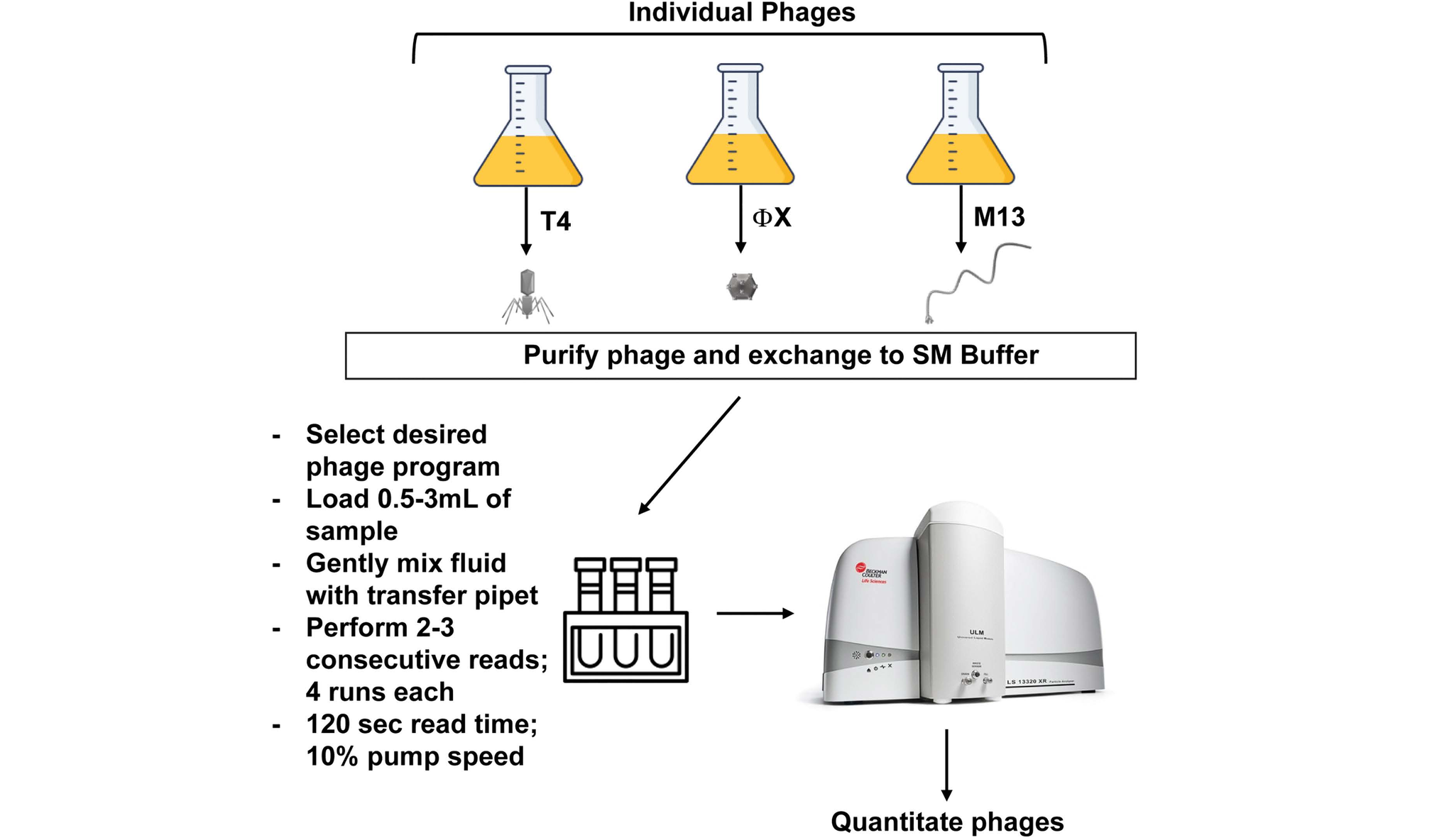
The method borrows an instrument from another field. Particle size analyzers, which measure tiny particles from the way they scatter light, have long been used to check powders and air quality. Newer models add a detector that compares how particles scatter light of several colors and polarizations, the directions in which light waves vibrate, making particles as small as phages visible. The researchers first showed that the analyzer could pick out phages in liquid, while plain buffer solution showed no particles. Because the instrument normally reports sizes rather than counts, a spreadsheet calculation was built to turn its readings into phage numbers, calibrated first against tiny latex beads and then fine-tuned using each phage’s measured size and counts from plaque tests.
To test the method, Dr. Carroll-Portillo’s team grew phages with very different shapes: a large virus with a head and spidery tail fibers, a small round one without a tail, and a long, thread-like one. Each sample was measured repeatedly on the analyzer and also counted with the plaque assay. Reported sizes varied from run to run, partly because phages are not perfect spheres and some clump together or break apart. Even so, the new counts fell within the accuracy range set against the plaque counts in the large majority of measurements, for every phage tested. “From these comparisons, we found that PhageFOTO was highly accurate for quantitation of all individual phages tested as compared to the plaque overlay assay,” explained Dr. Carroll-Portillo.
Results arrive in about an hour, without dyes or growing host bacteria, and this is the key advance. That helps when hosts are hard to grow or plaques are too faint to count, and it opens a path to samples holding phages of many different bacteria. Dr. Carroll-Portillo’s study also tested how dilute a sample can be: concentrated samples were detected reliably, very dilute ones were missed, and the detection limit was on par with established dye-based methods. “Development of a rapid, consistent, simple method for quantitation of phages in solution is desirable,” she said.
The work shows that an instrument built for chemistry and industry can become a fast, label-free phage counter. Because shape and clumping affect the readings, the calculation can be refined as more kinds of phages are measured, and all the data have been shared to help other laboratories. Next, Dr. Carroll-Portillo’s team plans to make the method more sensitive and to study mixtures of phages, a step towards counting phages in environmental samples. “PhageFOTO provides a rapid and reproducible method to quantitate phages from any phage-containing sample,” she explained.
About the researchers

Professor Henry Lin
Professor Henry Lin has been on the Faculty of the University of New Mexico since 2007. He is currently Professor of Medicine with Tenure. Dr. Lin received his MD from the State University of New York in 1982 after completing a 6-year B.S.-M.D. program.… He then completed an Internship and a Residency in Internal Medicine at the Harbor-UCLA Medical Center followed by a Fellowship in Gastroenterology at UCLA. Subsequent to his clinical training, he joined the Faculty of UCLA and USC before being recruited by University of New Mexico. His research interests are in nutrient regulation of gut motility and the role of resident gut bacteria and the gut microbiome-brain axis in health and disease. His bench-to-bedside research programs have been supported by the NIH, the VA, and the Department of Defense.

Dr. Amanda Carroll-Portillo
Dr. Amanda Carroll-Portillo is an Associate Research Professor at the University of New Mexico. She received her PhD in Microbiology from the University of Texas Health Science Center, San Antonio in 2004. She performed biomedical research in both academia and industry with focus on protein interactions, immunological responses, and novel detection methods. Currently, Dr.… Carroll-Portillo is investigating the functional role of bacteriophages within the gastrointestinal microbiome as it pertains to health and disease.

Cody Braun
Cody Braun began his research career as a field ecologist studying the spatial and social dynamics of collared lizards. He then taught General and Health Sciences Biology at Central New Mexico Community College for four years before joining the Lin lab in 2021.… Since that time he has worked on a variety of projects centered around small intestinal bacterial overgrowth, cellular responses to perturbation in gut homeostasis, and potential therapeutic interventions.
Journal reference
Carroll-Portillo A, Braun CA, Martinez E, Lin HC. Rapid bacteriophage quantification with a particle size analyzer combined with polarization intensity differential scattering (PIDS) detector. PLOS One. 2026; 21:e0343131. DOI: https://doi.org/10.1371/journal.pone.0343131