Feature Engineering + Physiologic MRI: Differentiating Glioblastomas from Brain Metastases
Dr. Sanjeev Chawla's team combined brain scan measurements of water movement and blood volume with machine learning to tell glioblastomas apart from cancers that spread to the brain.


Glioblastoma is an aggressive primary brain cancer, and brain metastases, which spread from cancers elsewhere in the body, represent the most common malignant tumors of the adult brain. Early differentiation is critical. While patients with glioblastoma are typically treated with surgery followed by chemotherapy and radiation therapy, those with suspected brain metastases and no history of systemic cancer require identification of the primary tumor site before any surgical, medical, or radiation therapy begins. On standard MRI scans, both tumors look very similar. Earlier attempts with advanced diffusion and perfusion MRI produced cut-off values that varied too much between studies, leaving radiologists/physicians unsure of how reliable they really are.
Dr. Sanjeev Chawla, Dr. Archith Rajan, Professor Steven Brem, Professor Laurie Loevner and Professor Suyash Mohan from the University of Pennsylvania, together with Dr. Seyyed Ali Hosseini, Dr. Stijn Servaes, Dr. Brandon Hall and Professor Pedro Rosa-Neto from McGill University and Dr. Sourav Bhaduri from The Chatterjee Group Centre for Research and Education in Science and Technology in India, built computer models that can distinguish between these two brain tumor types using advanced (physiologic) MRI scans. Their approach pairs measurements that reflect tumor biology with feature engineering, a technique that generates new variables and extracts new insights from existing data. This work has been published in the peer-reviewed journal, “Diagnostics”.
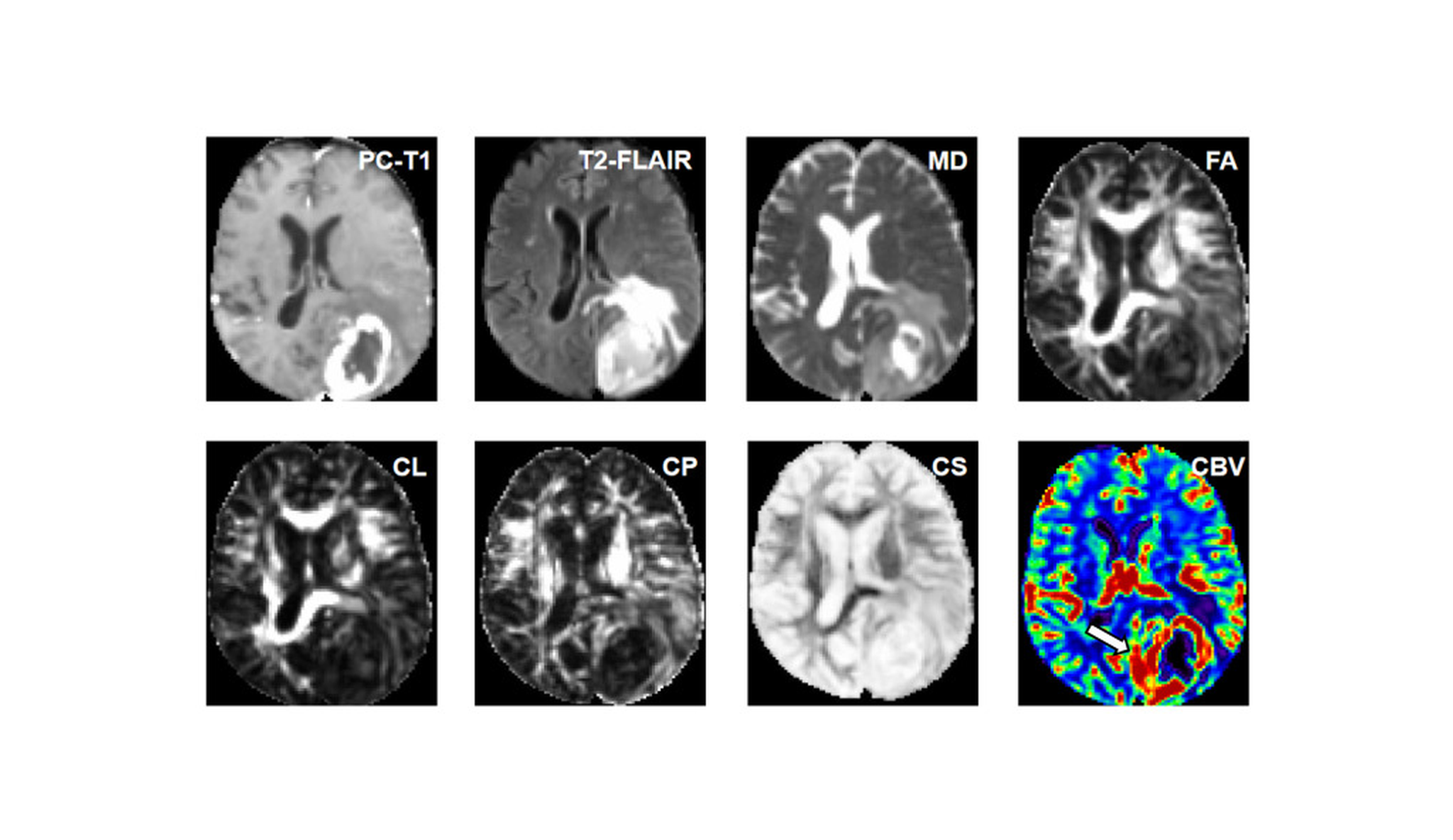
Prior to treatment, all patients each with a single brain tumor underwent an MRI on a 3T scanner, which also included an injection of contrast dye. The imaging protocol included structural scans to assess tumor size, brain location, and contrast enhancement patterns within the tumors. Additionally, advanced scans were performed such as diffusion tensor imaging, a scan that tracks how water molecules move through tissue, and perfusion imaging, which follows a dye through the blood vessels to estimate how much blood the tissue holds. The scans showed how freely and in which directions water moved, and how much blood was present, in the dye-enhanced part of the tumor and in a thin band of tissue surrounding it.
Dr. Chawla’s team isolated the most informative measurements and engineered new features by squaring variables and multiplying pairs. They evaluated various machine learning models using three configurations: the original metrics, the engineered features, and a combination of both. To ensure robust performance, each model underwent cross-validation, where it was repeatedly trained on a subset of the data and validated on the remaining portion.
The strongest single clues came from how strongly water movement favored certain directions inside the dye-enhanced tumor, and from water movement and blood volume in the surrounding band. Water moved significantly less freely around glioblastomas, which fits with cancer cells invading nearby tissue, whereas around metastases this zone mainly contains swelling with few or no cancer cells. “Probing the differences in imaging features from the regions around the tumor is an effective approach to differentiating glioblastomas from brain metastases,” explained Dr. Chawla.
The engineered and combined feature sets outperformed the original measurements across all performance metrics. The top-performing model, a random forest, utilized this combined dataset. When tested on patients held back from training data set, the model successfully classified most tumors. This yielded an improved overall diagnostic score compared to using either feature set in isolation, representing a substantial advancement over the baseline measurements. “Our findings indicate that feature engineering has a positive impact on the model’s performance,” said Dr. Chawla. This study is, to the team’s knowledge, the first to apply feature engineering to these measurements in brain tumor research.
Unlike earlier approaches built on many abstract shape and texture patterns, these models rely on measurements widely used in hospitals and linked directly to tumor biology. “The accurate and early diagnosis of glioblastomas versus single brain metastases provides a window of opportunity for reframing treatment strategies, enabling optimal and timely therapeutic interventions in these patients,” Dr. Chawla explained. Because the work involved a small group of patients at a single center and was not yet tested at other hospitals, larger studies across several hospitals with standardized scanning methods are the next step toward bringing the approach into routine clinical care.
About the researcher

Dr. Sanjeev Chawla
Dr. Sanjeev Chawla is a Research Associate Professor of Radiology at Perelman School of Medicine at the University of Pennsylvania. He is also a medical physicist certified by American Board of Medical Physics. He conducts a course on MRI Fundamentals (MPHY 6130) for graduate students during each Fall Semester at Penn Medicine. Dr.… Chawla received his master’s degree from Indian Institute of Technology, Delhi, India, and PhD degree in Radiology from Sanjay Gandhi Post-graduate Institute of Medical Sciences, Lucknow, India. Dr. Chawla’s research interests include implementation and application of MR imaging and spectroscopy methods for studying brain tumors, head & neck cancers and neurodegenerative diseases. He has authored more than 125 research/review articles and multiple book chapters. He has been awarded with Fulbright scholarship and research grants from NIH, ISMRM, ASHNR, PCPM, ITMAT, McCabe Foundation and Research Foundation of UPenn. Dr. Chawla was presented with Outstanding Researcher Award in Neuroradiology from Venus International Foundation and Distinguished Investigator Award from Academy of Radiology and Biomedical Research Imaging.
Journal reference
Hosseini SA, Servaes S, Hall B, Bhaduri S, Rajan A, Rosa-Neto P, Brem S, Loevner LA, Mohan S, Chawla S. Quantitative Physiologic MRI Combined with Feature Engineering for Developing Machine Learning-Based Prediction Models to Distinguish Glioblastomas from Single Brain Metastases. Diagnostics. 2024; 15(1):38. DOI: https://doi.org/10.3390/diagnostics15010038