Wired for Danger: Unveiling the Neural Secrets of Melanoma Metastasis
Professor Dominique Durand's team found that nerve activity recorded inside melanomas in mice could distinguish tumours that spread spontaneously from those with low metastatic potential.


Melanoma, though it accounts for only a small percentage of skin cancers, is responsible for the majority of skin cancer deaths. With the number of melanoma cases rising sharply in recent years, early detection is becoming increasingly critical. While current methods rely on visual examinations and advanced imaging techniques, there’s a growing interest in understanding the role of the nervous system in cancer development. Surprisingly, tumors have their own network of nerves, and these nerves might be key to unlocking new ways to predict how dangerous a melanoma can become.
Understanding the intricate behaviors of tumors has long been a focus for cancer researchers. Recent findings from Case Western Reserve University reveal a breakthrough in differentiating between melanomas based on their metastatic potential through neural recordings. The research, led by Dr. Grant McCallum and Professor Dominique Durand, along with Jay Shiralkar and Tiana Anthony, uncovers how the neural activity within tumors correlates with their metastatic behavior. Their study, published in PLOS ONE, marks a significant step toward early detection and treatment of melanoma.
The researchers conducted a series of experiments on mice to observe the neural patterns in metastasizing and non-metastasizing melanomas. Professor Durand explained the motivation behind the study: “Our goal was to determine if the bioelectric behavior of tumors could serve as an early indicator of their metastatic potential.”
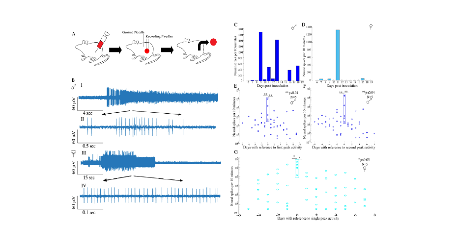
To explore the differences in neural activity between various types of melanomas, the research team used a combination of advanced neural recording techniques and bioluminescent imaging. They implanted electrodes in the tumors of mice to monitor neural spikes, providing a real-time look at the electrical activity within the tumors. This allowed the team to capture detailed neural patterns and correlate them with the tumors’ behavior over time. Daily recordings were taken to ensure that the data reflected the ongoing changes within the tumor environment.
The team discovered that melanomas with high metastatic potential exhibited significantly higher neural activity compared to those with low metastatic potential. This activity was particularly evident in the peaks observed in the neural recordings. Metastasizing tumors showed discontinuous trains of high neural activity, while non-metastasizing tumors exhibited minimal neural spikes. In simpler terms, the more aggressive tumors had more “active” nerves within them. The presence of sympathetic nerves played a crucial role in this activity. “Sympathectomy, or the chemical removal of sympathetic nerves, eliminated the peak neural activity in both sexes,” noted Dr McCallum, emphasizing the importance of these nerves in tumor progression. “Surprisingly, our research indicates that the brain is not only aware of the presence of a tumor but also establishes lines of communication to control it. The intricate neural activity we observed within melanomas suggests a complex interaction where the brain might influence tumor behavior and progression,” explained Professor Dominique Durand.
In addition to neural recordings, the researchers utilized bioluminescent imaging to track tumor growth and metastasis. By injecting a bioluminescent marker, they were able to visualize the tumors’ expansion and spread to other parts of the body, particularly the cranial area, which is a common site for melanoma metastasis. This method provided a comprehensive view of how the tumors developed and spread over time. The team observed that the neural activity peaks aligned closely with the onset of increased metastatic load, underscoring the potential of neural recordings as a predictive tool.
Additionally, the study found that tumors with low metastatic potential had significantly lower nerve density compared to their highly metastatic counterparts. This difference in nerve density further solidifies the link between neural activity and tumor aggressiveness.
The researchers believe that this study opens new avenues for early diagnosis and targeted therapy in melanoma treatment. “Our findings suggest that monitoring neural activity in tumors could provide a non-invasive method to predict their metastatic potential,” said Professor Durand. “This approach could lead to earlier interventions, potentially improving survival rates for melanoma patients.”
In conclusion, the study by Dr. McCallum, Professor Durand, and their colleagues presents compelling evidence that neural recordings can distinguish between melanomas based on their metastatic potential. This breakthrough not only enhances our understanding of tumor biology but also paves the way for innovative diagnostic tools in cancer treatment.
About the researchers

Dominique M. Durand
Dominique M. Durand is E.L. Linsedth Professor of Biomedical Engineering and Neurosciences and Director of the Neural Engineering Center at Case Western Reserve University in Cleveland, Ohio. He received an engineering degree from Ecole Nationale Superieure d'Electronique, Hydrolique, Informatique et Automatique de Toulouse, France in 1973. In 1974, he received a M.S.… degree in Biomedical Engineering from Case Reserve University in Cleveland OH., worked several years at the Addiction Research Foundation of Toronto, Canada and in 1982 received a Ph.D. in Electrical Engineering from the University of Toronto in the Institute of Biomedical Engineering. He received an NSF Young Investigator Presidential Award as well as the Diekhoff and Wittke awards for graduate and undergraduate teaching and the Mortar board top-prof awards at Case Western Reserve University. He is an IEEE Fellow and also Fellow of the American Institute for Medical and Biomedical Engineering and Fellow of the Institute of Physics. He serves on many editorial boards of peer-reviewed scientific journals. He is the founding editor of the Journal of Neural Engineering and served as editor-in-chief for 18 years. His research interests are in neural engineering and include computational neuroscience, neurophysiology and control of epilepsy, non-linear dynamics of neural systems, neural prostheses and applied magnetic and electrical field interactions with neural tissue. He has obtained funding for his research from the National Science Foundation, the National Institutes of Health and private foundations. He has published over 160 peer-reviewed articles and he has consulted for many biotechnology companies and foundations.

Grant A. McCallum
Grant A. McCallum received the Ph.D. degree in electrical engineering from Case Western Reserve University (CWRU) in 2011. He is currently a Research Assistant Professor in the Department of Biomedical Engineering at CWRU.… Prior to graduate studies, he worked for a total of nine years at Texas Instruments and nVidia Corporation as a Senior ASIC Design Engineer creating broadband access integrated circuits and graphics processors. His general research interests include the development of peripheral nerve interfaces, low-noise neural recording systems, and implantable biotelemetry devices.

Dr. Jay R Shiralkar
Dr. Jay R Shiralkar recently earned his PhD in Biomedical Engineering from Case Western Reserve University with guidance from eminent & distinguished Professor Dominique M Durand. His research focused on developing neural interfacing for solid tumors, with an emphasis on unraveling the role of autonomic nervous system in tumor physiology.… During his doctoral studies, Jay published papers in impactful journals, highlighting his contributions to the field of neural engineering & cancer biology and their applications in breast and melanoma tumors. Jay’s work has been recognized with awards, including the Swanger Fellowship Award from Case School of Engineering. He has also presented his findings at multiple international conferences, garnering attention for his novel approaches to solving complex biomedical problems. Beyond his research, Jay has demonstrated a strong commitment to mentorship and teaching, serving as a TA for several undergraduate courses and mentoring junior researchers in the lab. His dedication to education and innovation in biomedical engineering positions him as a promising emerging leader in the field. In his spare time, Jay enjoys volunteering in community health initiatives and exploring the latest advancements in medical technology. With a passion for improving patient outcomes through cutting-edge research, Dr. Jay R Shiralkar is poised to make significant contributions to the biomedical engineering and oncology community.
Journal reference
Shiralkar, J., Anthony, T., McCallum, G. A., & Durand, D. M. (2024). “Neural recordings can differentiate between spontaneously metastasizing melanomas and melanomas with low metastatic potential.” PLOS ONE, 19(2), e0297281. DOI: https://doi.org/10.1371/journal.pone.0297281